Changing the voltage of ordinary cells made frog embryos grow complete eyes, even on the gut
Michael Levin’s lab found a voltage signal that marks where a frog’s eyes will form, and showed that recreating it elsewhere grows eyes where none belong.
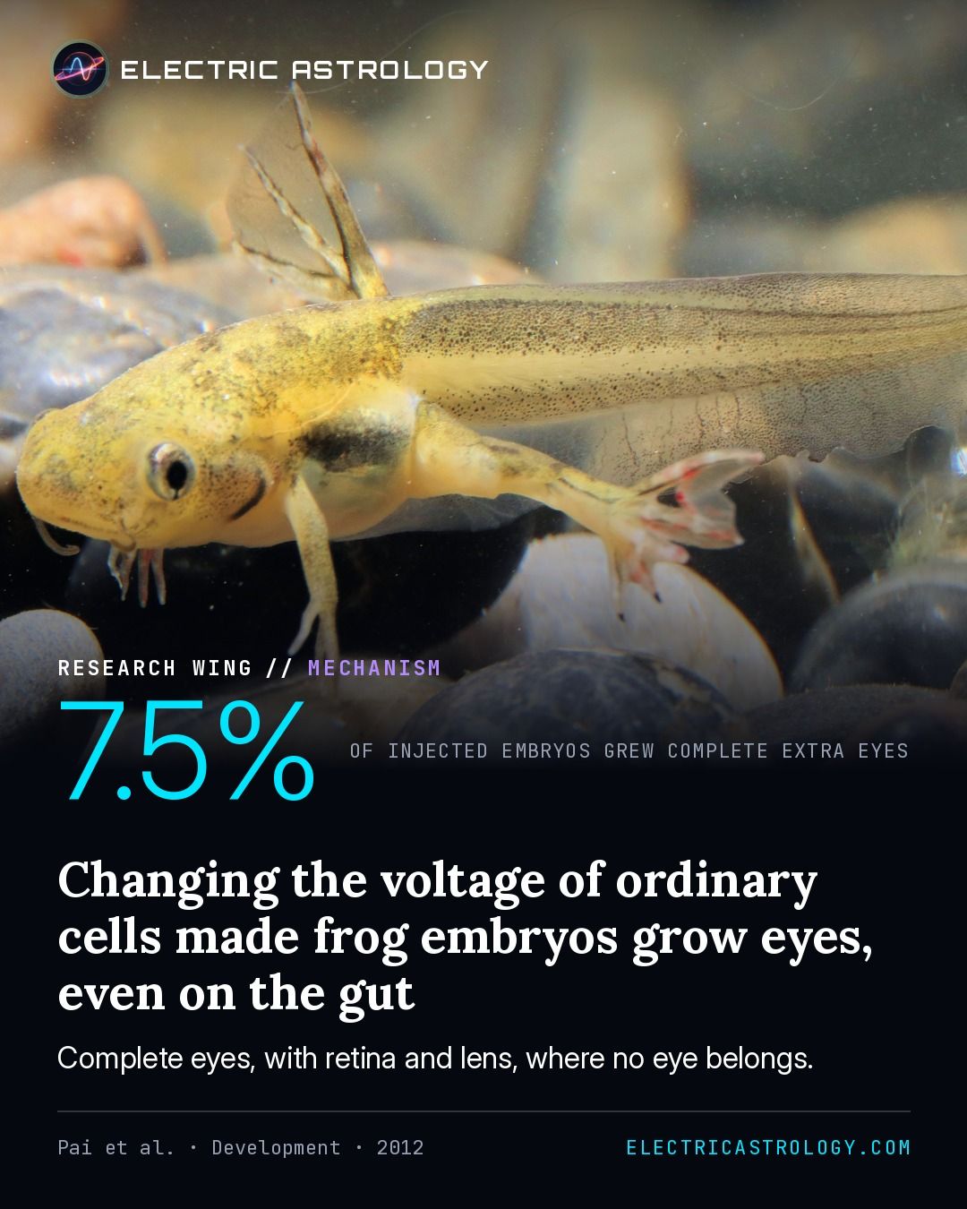
The short version
In frog embryos, the cells that will become the eyes sit at a more negative voltage than their neighbours. Researchers changed the voltage of other cells in the embryo by making them produce altered ion channels.
About one embryo in five grew extra eye tissue, and 7.5% grew complete eyes, with retina and lens. Extra eyes appeared even on the gut, and eye tissue as far back as the tail.
7.5%
Injected embryos that grew well-formed extra eyes, with pigmented epithelium, retina and lens. About 20% grew partial eye tissue.
The paper
Transmembrane voltage potential controls embryonic eye patterning in Xenopus laevis
Pai, V. P., Aw, S., Shomrat, T., Lemire, J. M., Levin, M. (2012). Transmembrane voltage potential controls embryonic eye patterning in Xenopus laevis. Development, 139(2), 313–323.
Checked against the paper’s full text on November 13, 2026.
What they measured
Using a voltage-reporting dye and direct electrical recordings, Vaibhav Pai and colleagues at Tufts mapped membrane voltage in embryos of the frog Xenopus laevis as the eyes are specified.
They then changed cell voltage by injecting mRNA for ion channels: channels that depolarize the eye-forming cells, and dominant-negative potassium-channel constructs that change the voltage of cells elsewhere in the embryo. Tadpoles were scored for eye defects and extra eyes.
What they found
From stage 17/18, two small patches in the front of the neural field, where the eyes will form, sit about 10 mV more negative than their neighbours (P = 0.0002).
Depolarizing those cells disrupted the normal eyes in 48% and 46% of embryos with two different channels, and restoring the voltage with outside chloride reduced the defects from 48% to 21%.
Changing the voltage of other cells produced extra eye tissue in about 20% of embryos and complete, well-formed eyes (pigmented epithelium, retina and lens) in 7.5%. Extra eyes appeared not only in the head but on the gut and in tail regions.
The authors conclude that membrane voltage is a crucial upstream signal in eye development, and that it can induce eyes far outside the region thought able to make them.
How it bears on the hypothesis
MechanismShows a working part of the circuit: a receiver in the body, a current in space, a field that couples them.
An eye is usually explained by genes switched on in the right place. Here the switch is electrical: give ordinary cells the right voltage and they build an eye, wherever they are.
The voltage works as a code, not as raw power. The authors found a narrow window of voltages compatible with eyes, which fits their observation that only some injected cells formed eye tissue.
The authors’ own caution: The authors could not fully rescue the depolarized eyes without disturbing other voltage-sensitive systems, and say full understanding would need extensive profiling of the native ion channels. (Our note: the paper does not test whether the extra eyes could see.)
On Electric Astrology
More on: Is the body’s shape stored in voltage?
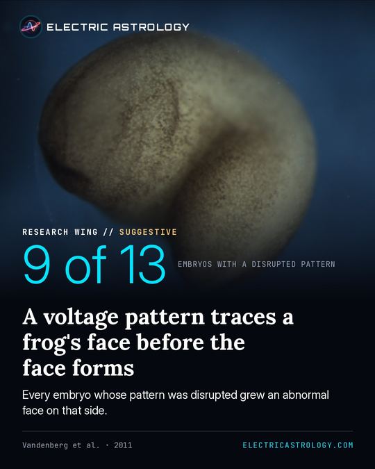 A voltage pattern traces a frog's face before the face formsVandenberg et al. · 2011 · Developmental Dynamics
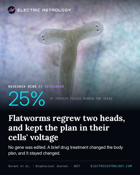 Flatworms regrew two heads, and kept the plan in their cells' voltageDurant et al. · 2017 · Biophysical Journal
 Mutations in one potassium-channel gene change the heart's rhythm and the facePlaster et al. · 2001 · Cell